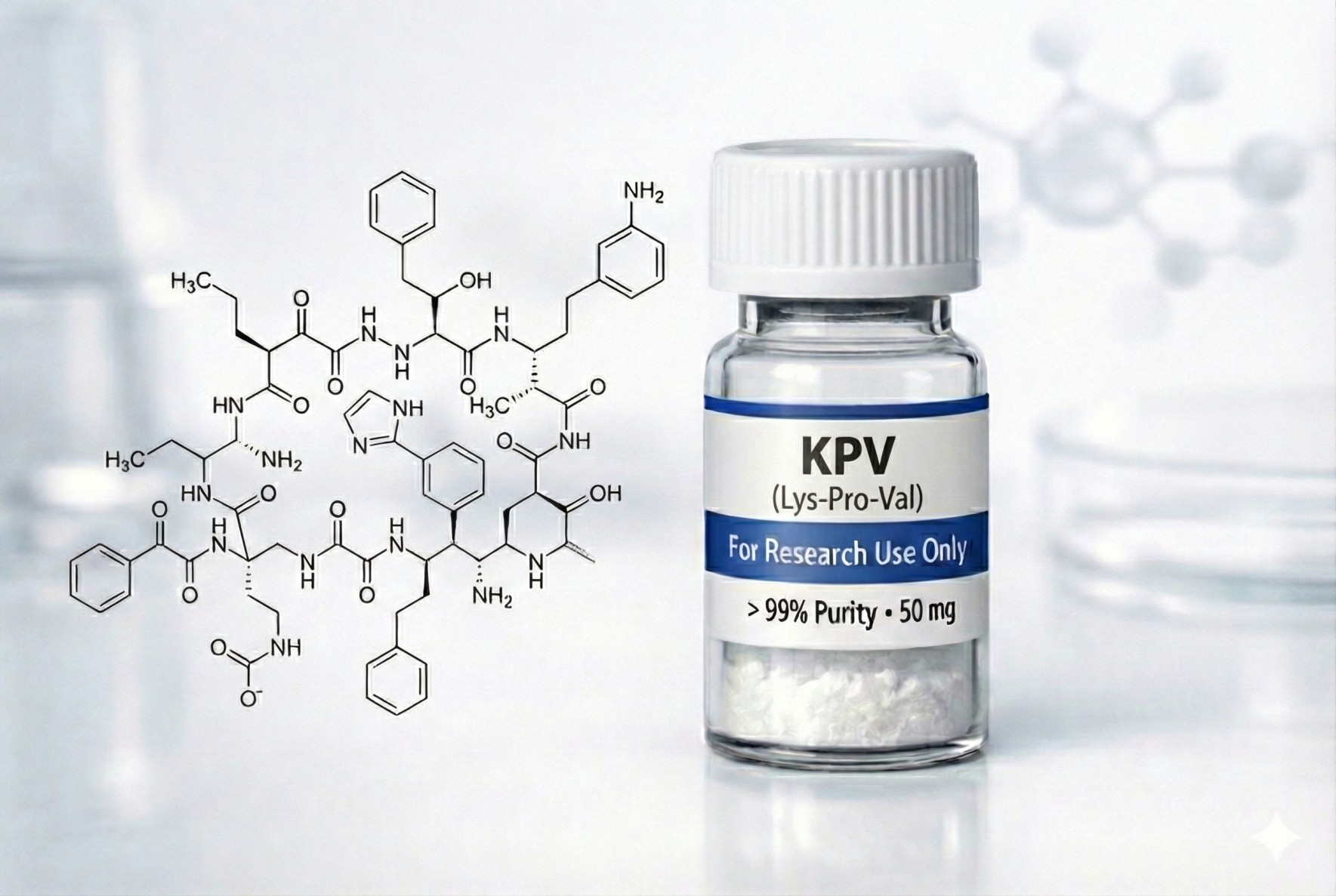
KPV is a naturally occurring tripeptide with the amino acid sequence Lysine-Proline-Valine (Lys-Pro-Val) and molecular weight of 369 Da.¹ ² ³ The peptide represents the three C-terminal amino acids of α-melanocyte-stimulating hormone (α-MSH), a 13-amino acid neuropeptide hormone (Ac-Ser-Tyr-Ser-Met-Glu-His-Phe-Arg-Trp-Gly-Lys-Pro-Val-NH₂) that exerts potent anti-inflammatory effects when administered systemically or locally.¹ ² α-MSH is derived from the precursor proopiomelanocortin (POMC) through post-translational proteolytic processing.¹ ²
**Historical Context:**¹ ²
α-MSH has been recognized since the late 1980s as having potent anti-inflammatory properties extending beyond its classical role in melanogenesis (skin pigmentation).¹ ² Research by Luger, Lipton, and colleagues demonstrated that α-MSH could suppress inflammation in diverse experimental models including contact dermatitis, inflammatory bowel disease, arthritis, asthma, and uveitis.¹ ² However, clinical development of α-MSH as an anti-inflammatory agent faced limitations due to its melanotropic effects—binding to melanocortin receptors (particularly MC-1R) on melanocytes causes increased melanin production and skin darkening, an undesirable cosmetic side effect for chronic systemic use.¹ ²
Research by Hiltz and Lipton in 1989 first demonstrated that the C-terminal tripeptide fragment KPV retained anti-inflammatory activity comparable to or even exceeding full-length α-MSH.² ⁴ Subsequent studies confirmed that KPV possesses “a similar or even more pronounced anti-inflammatory activity as full-length α-MSH” while eliminating melanotropic effects.² This discovery opened the possibility of developing KPV as a targeted anti-inflammatory agent without cosmetic complications.
**Unique Mechanism—PepT1 Transporter, Not Melanocortin Receptors:**³
Unlike α-MSH, which mediates anti-inflammatory effects primarily through melanocortin receptor (MC-1R, MC-3R) activation and cyclic AMP elevation, KPV operates via an entirely distinct mechanism:³ ⁴
Does Not Bind Melanocortin Receptors: Multiple studies confirmed that KPV does not bind to MC-1R, MC-3R, or MC-5R, does not increase intracellular cyclic AMP levels, and does not stimulate melanocytes.² ³ ⁴ This explains why KPV lacks pigmentary effects despite retaining anti-inflammatory properties.
PepT1-Mediated Cellular Uptake: The landmark 2008 study by Dalmasso et al. at Emory University definitively established that KPV’s anti-inflammatory mechanism depends on cellular uptake via the PepT1 transporter (SLC15A1), an H⁺-coupled oligopeptide transporter that normally transports dietary di- and tripeptides in the small intestine.³
Strategic Expression Pattern: PepT1 is normally expressed on the apical (luminal) membrane of small intestinal enterocytes but is virtually absent from normal colon.³ Critically, PepT1 is dramatically upregulated in inflamed colonic epithelial cells during inflammatory bowel disease (IBD), providing a disease-specific delivery mechanism.³ Additionally, immune cells including macrophages and T lymphocytes (Jurkat cells) express functional PepT1, allowing KPV to target both epithelial and immune components of intestinal inflammation.³
High Affinity Transport: PepT1 transports KPV with remarkably high affinity (Km ~160 μM in Caco2-BBE intestinal epithelial cells, Km ~700 μM in Jurkat T cells)—among the lowest Km values reported for PepT1 substrates.³ For comparison, the commonly used PepT1 substrate Gly-Sar has Km ≥1 mM.³ This high affinity allows efficient cellular uptake at low nanomolar concentrations.
Intracellular Anti-Inflammatory Action: Once transported into cells, KPV accumulates intracellularly and directly inhibits inflammatory signaling cascades including NF-κB and MAPK pathways, reducing pro-inflammatory cytokine production (IL-8, IL-1β, IL-6, TNF-α).³ The peptide does not act at the cell surface but must enter cells to exert effects.³
Pharmacological Characteristics:
Very Short Systemic Half-Life: As a small unmodified tripeptide, KPV is rapidly degraded by peptidases in serum and tissues, resulting in extremely short systemic half-life (likely minutes).² ³ This necessitates either frequent dosing, local administration, or advanced delivery systems.
Oral Bioavailability via PepT1: Unlike most peptides that are rapidly degraded in the gastrointestinal tract, KPV can be absorbed orally via PepT1 expressed on small intestinal enterocytes.³ Preclinical studies successfully demonstrated efficacy using oral administration (added to drinking water), with KPV reaching and acting on inflamed colonic tissues.³
Local Action at Inflamed Sites: KPV’s dependence on PepT1—upregulated specifically in inflamed tissues—provides inherent targeting to sites of active inflammation while sparing normal tissues.³ This tissue-selective mechanism theoretically minimizes systemic side effects.
Nanoparticle Delivery Enhancement: Advanced drug delivery systems using hyaluronic acid-functionalized nanoparticles encapsulated in pH-sensitive hydrogels achieved 12,000-fold enhancement in anti-inflammatory potency compared to free KPV solution, allowing therapeutic effects at nanomolar concentrations.⁵
Routes of Administration: Preclinical studies employed oral (drinking water), intraperitoneal injection, topical (cream/ointment), and advanced nanoparticle formulations.² ³ ⁵
Regulatory and Legal Status:
**FDA Category 2 Classification (September 2023):**⁶ ⁷ ⁸
The FDA placed KPV on the Category 2 Bulk Drug Substances List, designating it as a substance that “raises significant safety concerns.”⁶ ⁷ The agency specifically cited “lack of sufficient safety data for human use” among the grounds for this classification.⁶ ⁷ ⁸ This Category 2 designation effectively prohibits compounding pharmacies from using KPV in compounded medications under FDA regulations.⁶ ⁷ ⁸
**Zero Human Clinical Trials:**⁶ ⁷
“There have been no human trials on KPV to date.”⁶ Despite over 15 years of preclinical research, KPV has never been evaluated in human subjects for safety or efficacy in any condition.⁶ ⁷
**Never FDA-Approved:**⁶ ⁷
KPV is not an FDA-approved drug and is not approved in any country surveyed.⁶ ⁷ It has never undergone the regulatory process required for pharmaceutical approval.
**Current Legal Status:**⁶ ⁷
KPV is not a DEA scheduled substance, so possession is not illegal.⁶ However, the FDA Category 2 designation prohibits its use in compounded medications.⁶ ⁷ ⁸ The peptide remains legally sold as “research chemicals” or “dietary supplements”—classifications not subject to FDA regulations for pharmaceutical quality, safety, and efficacy.⁶ ⁷ This creates a gray-area legal status where cash-based medical clinics offer KPV treatment despite zero human safety data and explicit FDA safety concerns.⁶ ⁷
How It Works
PepT1-Mediated Cellular Uptake and Intracellular Action
KPV’s anti-inflammatory mechanism fundamentally differs from its parent molecule α-MSH and represents a novel approach to targeting inflammation.³
Step 1: PepT1 Transport into Cells³
KPV is transported across cell membranes via the PepT1 transporter (SLC15A1), an H⁺-coupled oligopeptide cotransporter that simultaneously transports peptides and protons.³ PepT1 normally functions in dietary peptide absorption in the small intestine but is dramatically upregulated in inflamed colonic epithelial cells during IBD and expressed by immune cells (macrophages, T lymphocytes) infiltrating inflamed tissues.³
Competitive Inhibition Studies: When Glycine-Leucine (Gly-Leu, a standard PepT1 substrate) was added to cells, it completely blocked KPV’s anti-inflammatory effects by competing for PepT1 transport.³ This definitively proved KPV requires cellular uptake via PepT1 to function.³
Cell Type Specificity: In HT29-Cl.19A human colonic cells that do not express PepT1, KPV had no anti-inflammatory effect despite these cells expressing functional IL-1β receptors and showing robust inflammatory responses to IL-1β.³ However, when HT29-Cl.19A cells were transfected with PepT1, KPV regained anti-inflammatory activity.³ This genetic evidence confirmed PepT1 as essential for KPV’s mechanism.³
High-Affinity Transport: Kinetic uptake experiments using radiolab
eled [³H]KPV demonstrated PepT1-mediated transport with Km ~160 μM in intestinal epithelial cells and ~700 μM in T cells—among the highest affinities (lowest Km values) reported for PepT1 substrates.³
Step 2: Intracellular Accumulation and NF-κB Inhibition³
Once inside cells, KPV accumulates intracellularly and directly inhibits the NF-κB (nuclear factor-kappa B) signaling pathway, a master regulator of inflammatory gene expression:³
IκB-α Degradation Inhibition: In intestinal epithelial cells (Caco2-BBE) stimulated with IL-1β, KPV significantly reduced degradation of IκB-α (the inhibitory protein that sequesters NF-κB in the cytoplasm).³ IL-1β normally causes rapid IκB-α degradation within 20 minutes, allowing NF-κB to translocate to the nucleus.³ In the presence of KPV, IκB-α degradation was reduced, and baseline IκB-α levels recovered much faster (90 minutes vs. 180 minutes), indicating that “KPV delays NF-κB activation and also shortened the delay of IκB-α recovery.”³
IκB-α Phosphorylation Blockade: KPV prevented phosphorylation of IκB-α at 45 minutes post-IL-1β stimulation, further confirming suppression of the NF-κB activation cascade.³
NF-κB Transcriptional Activity: Using NF-κB-dependent luciferase reporter assays, KPV at 10 nM concentration significantly decreased IL-1β-induced NF-κB transcriptional activity by ~6-fold.³ This directly demonstrates that KPV suppresses NF-κB-driven gene expression.³
EMSA Confirmation: Electrophoretic mobility shift assays (EMSA) confirmed that KPV reduces NF-κB DNA binding activity.³
Step 3: MAPK Pathway Inhibition³
KPV strongly inhibits activation of mitogen-activated protein kinase (MAPK) inflammatory signaling pathways:³
IL-1β normally induces rapid phosphorylation (activation) of three major MAPK family members: ERK1/2, JNK, and p38.³ Co-treatment with KPV (10 nM) “strongly decreased IL-1β-induced MAPK phosphorylation and, therefore, their activation.”³ This multi-pathway inhibition contributes to KPV’s broad anti-inflammatory effects.³
Step 4: Pro-Inflammatory Cytokine Suppression³
By inhibiting NF-κB and MAPK pathways, KPV reduces transcription and secretion of pro-inflammatory cytokines:³
IL-8 Reduction (Intestinal Epithelial Cells): IL-1β induced ~200-fold increase in IL-8 mRNA in Caco2-BBE cells.³ KPV co-treatment reduced this increase by ~35%.³ IL-8 protein secretion into culture medium was similarly decreased.³ IL-8 is a key neutrophil chemoattractant, so reducing IL-8 decreases immune cell recruitment to inflamed tissues.³
Broad Cytokine Suppression (Multiple Cell Types): KPV reduces production of IL-1β, IL-6, IL-12, TNF-α, and IFN-γ across intestinal epithelial cells, macrophages, and T lymphocytes.² ³ Importantly, KPV did not reduce IL-10 (anti-inflammatory cytokine), suggesting selective suppression of pro-inflammatory mediators rather than global immunosuppression.³
Step 5: Immune Cell Function Modulation³
T Lymphocytes (Jurkat Cells): KPV reduced TNF-α-induced IκB-α degradation and IL-8 mRNA expression in human T cells, demonstrating effects on adaptive immune responses.³
Macrophages: KPV suppresses LPS-induced inflammatory responses in macrophages, reducing pro-inflammatory cytokine production.² ³
Neutrophil Recruitment: By reducing IL-8 and other chemokines, KPV inhibits neutrophil recruitment and transmigration across the intestinal epithelium, decreasing tissue damage caused by infiltrating immune cells.³
Additional Mechanisms
**IL-1β Receptor Antagonism:**¹ ²
A stereochemical analog of KPV (K(D)PT, where valine is replaced with threonine) corresponds to amino acids 193-195 of IL-1β.¹ ² During IL-1β degradation, this KPT loop is revealed on the protein surface and can interact with IL-1 receptor type I (IL1RI), exerting antagonistic activity and contributing to terminating IL-1β-mediated inflammation.² While KPV itself was not found to act as an IL-1β receptor antagonist in the Dalmasso study (it had no effect in cells lacking PepT1 despite functional IL-1β receptors),³ this mechanism may contribute to the effects of KPV analogs.
**Antimicrobial Activity:**² ⁹
Beyond anti-inflammatory effects, KPV and α-MSH demonstrate antimicrobial activity against pathogens including Staphylococcus aureus and Candida albicans.² ⁹ The candidacidal activity is believed to be mediated by increased cellular cyclic AMP, though this appears distinct from the melanocortin receptor-independent anti-inflammatory mechanism.² This dual anti-inflammatory and antimicrobial activity suggests KPV may reduce both inflammation and infection risk—an advantage over traditional immunosuppressive agents.²
Research Evidence
Preclinical Studies—Inflammatory Bowel Disease
Dalmasso G et al., 2008 – PepT1-Mediated Tripeptide KPV Uptake Reduces Intestinal Inflammation
Design: Comprehensive in vitro and in vivo study examining KPV’s anti-inflammatory mechanism and efficacy in experimental colitis models.³
Published: Gastroenterology, 2008 Jan;134(1):166-178. (Top-tier gastroenterology journal)³
**In Vitro Studies:**³
Intestinal Epithelial Cells (Caco2-BBE):
- KPV (10 nM) significantly reduced IL-1β-induced NF-κB luciferase activity (~6-fold reduction)
- Reduced IκB-α degradation and phosphorylation
- Strongly decreased ERK1/2, JNK, and p38 MAPK phosphorylation
- Reduced IL-8 mRNA expression by ~35% and IL-8 protein secretion
Mechanism Confirmation:
- Gly-Leu (PepT1 substrate) completely reversed KPV’s anti-inflammatory effects, proving PepT1-dependence
- KPV had no effect in HT29-Cl.19A cells lacking PepT1 despite functional IL-1β receptors
- Transfecting HT29-Cl.19A cells with PepT1 restored KPV’s anti-inflammatory activity
- α-MSH had no anti-inflammatory effect in these systems despite melanocortin receptor expression
PepT1 Transport Kinetics:
- Km ~160 μM for KPV in Caco2-BBE cells (high affinity)
- 100 μM KPV inhibited Gly-Sar uptake by ~45% vs. ~25% for Gly-Leu, indicating higher PepT1 affinity for KPV
- Nanomolar concentrations of [³H]KPV efficiently transported by PepT1
T Lymphocytes (Jurkat Cells):
- KPV reduced TNF-α-induced IκB-α degradation at 15 minutes
- Reduced IL-8 mRNA expression by ~5-fold at 6 hours
- Km ~700 μM for KPV transport in Jurkat cells
- First demonstration that human T cells express functional PepT1 capable of transporting KPV
**In Vivo Studies—DSS-Induced Colitis (N=10 mice/group):**³
Model: Dextran sulfate sodium (DSS) 3% in drinking water for 8 days; KPV 100 μM added to drinking water
Results:
- Body Weight: DSS caused characteristic weight loss starting day 4; KPV significantly reduced weight loss at day 8 vs. DSS alone
- Myeloperoxidase (MPO) Activity: DSS-induced increase in MPO (neutrophil infiltration marker) was decreased by ~50% with KPV treatment
- Histology: DSS caused cell wall damage, interstitial edema, and increased inflammatory cell infiltration in lamina propria; mice receiving DSS + KPV showed “markedly reduced intestinal inflammation”
- Colon Weight and Length: KPV prevented DSS-induced increase in colon weight and decrease in colon length
- Pro-Inflammatory Cytokines (RT-PCR):
- IL-6 mRNA: significantly reduced by KPV (p<0.05)
- IL-12 mRNA: significantly reduced by KPV (p<0.05)
- IL-1β mRNA: reduced (trend)
- IFN-γ mRNA: reduced (trend)
- IL-10 mRNA: no change (anti-inflammatory cytokine unaffected)
- KPV Alone: Had no effect on basal MPO levels or inflammatory parameters in normal mice, indicating KPV specifically targets inflammation without affecting homeostasis
**In Vivo Studies—TNBS-Induced Colitis (N=10 mice/group):**³
Model: Trinitrobenzene sulfonic acid (TNBS) 150 mg/kg colonic injection; KPV 100 μM in drinking water; assessment at 48 hours
Results:
- Body Weight: KPV significantly reduced weight loss at days 1 and 2 vs. TNBS alone
- MPO Activity: TNBS-induced MPO increase was inhibited by ~30% with KPV
- Colon Length: KPV prevented TNBS-induced decrease in colon length
- Pro-Inflammatory Cytokines (RT-PCR):
- IL-1β mRNA: significantly reduced (p<0.05)
- IL-6 mRNA: significantly reduced (p<0.05)
- TNF-α mRNA: significantly reduced (p<0.05)
- IFN-γ mRNA: significantly reduced (p<0.05)
**Significance:**³
This landmark study:³
- Definitively established KPV’s mechanism: PepT1-mediated cellular uptake followed by intracellular NF-κB and MAPK inhibition
- First report of KPV-mediated reduction of colitis: Demonstrated efficacy in two distinct experimental IBD models
- Confirmed oral bioavailability: Showed that orally administered KPV can reach inflamed colon and exert therapeutic effects
- Identified PepT1 as therapeutic target: Upregulation of colonic PepT1 during IBD provides disease-specific delivery mechanism
- Published in top-tier journal: Gastroenterology is among the most prestigious journals in gastroenterology research
Preclinical Studies—Contact Dermatitis
Getting SJ et al., 2003 – Dissection of the anti-inflammatory effect of the core and C-terminal (KPV) alpha-melanocyte-stimulating hormone peptides
Design: Study comparing anti-inflammatory effects of full-length α-MSH vs. KPV in contact dermatitis model.⁴
Model: Oxazolone-induced contact hypersensitivity in mice
**Results:**⁴
- Both α-MSH and KPV, administered intravenously or topically, suppressed contact dermatitis reactions
- KPV produced anti-inflammatory effects comparable to or exceeding α-MSH
- KPV induced hapten-specific tolerance when applied topically
**Mechanism Differentiation:**⁴
- α-MSH’s effects were melanocortin receptor-dependent (particularly MC-1R)
- KPV’s effects were melanocortin receptor-independent
- KPV lacked melanotropic activity (no skin pigmentation changes)
Significance: Established that KPV retains full anti-inflammatory efficacy of α-MSH without melanocortin receptor binding or pigmentary side effects.⁴
Advanced Drug Delivery—Nanoparticle Systems
Xiao B et al., 2017 – Orally Targeted Delivery of Tripeptide KPV via Hyaluronic Acid-Functionalized Nanoparticles Efficiently Alleviates Ulcerative Colitis
Design: Development and evaluation of hyaluronic acid (HA)-functionalized KPV-loaded nanoparticles (HA-KPV-NPs) embedded in chitosan/alginate hydrogel for oral delivery to inflamed colon.⁵
**Rationale:**⁵
- Free KPV requires high concentrations (micromolar) for therapeutic effect
- Nanoparticle targeting to inflamed mucosa could dramatically enhance efficacy
- HA binds to CD44 receptors overexpressed on inflamed colonic epithelium and immune cells
- Hydrogel protects NPs during gastric/small intestinal transit and releases NPs at colonic pH
**Nanoparticle Characteristics:**⁵
- Poly(lactic-co-glycolic acid) (PLGA) core loaded with KPV
- Surface-functionalized with hyaluronic acid
- Encapsulated in pH-sensitive chitosan/alginate hydrogel
- Collapses at colonic pH (~7) to release HA-KPV-NPs
**In Vitro Results:**⁵
- HA-KPV-NPs showed abundant internalization into colonic epithelial cells and macrophages
- Penetrated deeply into inflamed tissue
- Enhanced cellular uptake compared to non-functionalized KPV-NPs
- Accelerated mucosal healing and alleviated inflammation in vitro
**In Vivo Results (DSS-Induced Colitis in Mice):**⁵
- Efficacy comparison:
- HA-KPV-NP/hydrogel system: highly effective at preventing mucosa damage
- Non-functionalized KPV-NP/hydrogel: moderately effective
- Free KPV solution: minimal effect at same total KPV dose
- 12,000-fold enhancement: HA-KPV-NPs achieved therapeutic effects at KPV concentrations 12,000-fold lower than free KPV solution
- TNF-α downregulation: Much stronger suppression with HA-KPV-NPs vs. KPV-NPs
- Safety: HA-KPV-NPs were non-toxic and biocompatible with intestinal cells
Significance: Demonstrated that advanced drug delivery systems can dramatically enhance KPV’s therapeutic potential, achieving efficacy at nanomolar concentrations and providing proof-of-concept for clinical translation.⁵
Broader Preclinical Evidence
Luger TA & Brzoska T, 2007 – α-MSH related peptides: a new class of anti-inflammatory and immunomodulating drugs
Design: Comprehensive review of α-MSH and KPV preclinical evidence across multiple inflammatory disease models.²
**Animal Models Demonstrating KPV Efficacy:**²
- Contact dermatitis: KPV applied IV or topically suppressed both sensitization and elicitation phases; induced hapten-specific tolerance
- Inflammatory bowel disease: KPV significantly reduced DSS- and TNBS-induced colitis
- Allergic asthma: α-MSH/KPV inhibited allergic airway inflammation; reduced IL-4 and IL-13 in bronchoalveolar lavage fluid
- Arthritis: α-MSH/KPV attenuated adjuvant-induced experimental arthritis; similarly effective as prednisolone without weight loss
- Uveitis: α-MSH/KPV suppressed experimental autoimmune uveitis and endotoxin-induced uveitis
- Cutaneous vasculitis: Single injection suppressed LPS-induced Shwartzman reaction
**Mechanisms Across Models:**²
- Suppression of NF-κB activation
- Reduced expression of adhesion molecules (ICAM-1, P-selectin)
- Decreased pro-inflammatory cytokines (IL-1β, IL-6, TNF-α, IFN-γ)
- Increased anti-inflammatory IL-10 in some models
- Inhibition of immune cell migration and recruitment
- Induction of regulatory T cells (in some models)
**Safety Profile:**²
- α-MSH and KPV were well tolerated in animal studies
- No significant adverse effects reported across multiple models
- KPV lacks melanotropic effects (no skin pigmentation) unlike α-MSH
- Activation of NF-κB was “never fully suppressed, but mostly only reduced,” suggesting KPV modulates rather than completely blocks inflammatory pathways²
- In absence of inflammation, KPV’s immunosuppressive effects were “usually weak or absent,” indicating selective action on active inflammation²
Significance: This comprehensive review established KPV as a broad-spectrum anti-inflammatory agent effective across diverse experimental models with favorable preclinical safety profile.²
Current Status & Considerations
Regulatory Status and Clinical Development
**FDA Category 2 Designation (September 2023):**⁶ ⁷ ⁸
The FDA placed KPV on the Category 2 Bulk Drug Substances List, designating it as raising “significant safety concerns.”⁶ ⁷ ⁸ The agency specifically cited “lack of sufficient safety data for human use” as grounds for this classification.⁶ ⁷ This designation effectively prohibits compounding pharmacies from using KPV in compounded medications under FDA regulations.⁶ ⁷ ⁸
**Zero Human Clinical Trials:**⁶ ⁷
Despite robust preclinical evidence dating back to the 1980s-1990s and mechanistic studies through 2017:
- “There have been no human trials on KPV to date.”⁶
- Zero Phase I safety trials in healthy volunteers
- Zero Phase II efficacy trials in any patient population
- Zero published case reports or case series
- Complete absence of human safety data
- Unknown human pharmacokinetics
- Unknown dosing requirements for any human condition
**Never FDA-Approved:**⁶ ⁷
KPV has never been FDA-approved for any indication and is not approved in any country surveyed.⁶ ⁷ It has never undergone the regulatory approval process required for pharmaceutical drugs.
**Current Legal Status:**⁶ ⁷
KPV is not a DEA scheduled substance, so possession is not illegal.⁶ However, the FDA Category 2 designation prohibits its use in compounded medications.⁶ ⁷ ⁸ Despite this, KPV remains available through:⁶ ⁷
- Unregulated “research chemical” suppliers
- Cash-based medical clinics operating outside evidence-based medicine
- Online vendors marketing as “dietary supplements” (not subject to FDA pharmaceutical regulations)
This creates a problematic legal gray zone where clinics offer KPV treatment despite zero human safety data and explicit FDA safety concerns.⁶ ⁷
Evidence Base Summary
**Preclinical Evidence:**² ³ ⁴ ⁵
The preclinical evidence base for KPV is substantial and scientifically rigorous:
Mechanism of Action:
- Definitively established via genetic validation (PepT1 knockout/transfection studies)³
- Confirmed across multiple cell types (intestinal epithelial cells, T lymphocytes, macrophages)³
- Mechanistic pathway clearly delineated (PepT1 uptake → intracellular NF-κB/MAPK inhibition)³
- Distinguished from parent molecule α-MSH (melanocortin receptor-independent)⁴
Animal Model Efficacy:
- Demonstrated across 6+ distinct inflammatory disease models²
- Multiple independent research groups (Emory University, University of Muenster, William Harvey Research Institute)² ³ ⁴
- Published in top-tier peer-reviewed journals (Gastroenterology, Annals of Rheumatic Diseases, J Pharmacol Exp Ther)² ³ ⁴
- Consistent findings using different routes of administration (oral, IV, topical)² ³ ⁴
- Both prevention and treatment paradigms evaluated³
Advanced Delivery Systems:
- Nanoparticle formulations achieved 12,000-fold potency enhancement⁵
- Proof-of-concept for clinical translation⁵
**Human Evidence:**⁶ ⁷
**Total human data: Zero studies, zero subjects, zero safety information.**⁶ ⁷
This represents an extraordinary disconnect—robust preclinical development over 15+ years with zero translation to human testing.⁶ ⁷
Critical Evidence Gaps
**Complete Absence of Human Safety Data:**⁶ ⁷
- Unknown human pharmacokinetics (absorption, distribution, metabolism, excretion)
- Unknown human dosing requirements
- Unknown acute toxicity profile in humans
- Unknown allergic/hypersensitivity potential
- Unknown long-term safety (chronic dosing effects)
- Unknown effects on human immune function
- Unknown drug-drug interactions
- Unknown effects in special populations (elderly, pregnant/lactating, pediatric, immunocompromised)
- Unknown systemic exposure with oral vs. topical administration
**Efficacy Questions:**⁶ ⁷
- No evidence KPV’s impressive preclinical efficacy translates to human disease
- Unknown whether human IBD responds similarly to mouse models
- Unknown optimal dosing, frequency, duration for any human condition
- Unknown which patient populations most likely to benefit
- No biomarkers validated for monitoring treatment response
**Manufacturing and Quality:**⁶ ⁷
- No pharmaceutical-grade manufacturing standards established
- Products from “research chemical” suppliers lack quality control, purity testing, sterility verification
- Unknown contamination/impurity risks with unregulated products
- No standardized formulations developed
Theoretical Safety Considerations
**Based on Mechanism and Preclinical Data:**² ³
Favorable Theoretical Safety Profile:
- Selective action: PepT1-mediated mechanism targets inflamed tissues (where PepT1 is upregulated) while sparing normal tissues³
- Modulation vs. suppression: KPV reduces but does not completely block NF-κB activation, maintaining baseline immune function²
- Context-dependent: In absence of inflammation, KPV shows minimal immunosuppressive effects²
- No melanotropic effects: Unlike α-MSH, KPV does not affect skin pigmentation²
- Antimicrobial activity: Dual anti-inflammatory and antimicrobial properties may reduce infection risk vs. traditional immunosuppressants²
**Preclinical Safety:**² ³
- Well tolerated across multiple animal models
- No significant adverse effects reported in published studies
- Effective doses did not cause weight loss (unlike corticosteroids)²
- No apparent organ toxicity in animal studies
Theoretical Concerns (Extrapolated from Mechanism):
Immunosuppression Risk: While KPV modulates rather than blocks inflammation,² chronic suppression of NF-κB and MAPK pathways could theoretically impair host defense against infections or tumor surveillance. However, no such effects were observed in animal studies.²
GI-Specific Concerns: High PepT1 expression in small intestine means KPV could affect normal small intestinal cells, not just inflamed colon.³ Effects on normal intestinal function unknown.
Peptide Immunogenicity: Chronic exposure to peptides can elicit antibody responses. KPV’s small size (tripeptide) may reduce immunogenicity risk, but human data are absent.
Manufacturing Impurities: Unregulated products may contain bacterial endotoxins, peptide degradation products, or synthesis byproducts with unknown toxicity.⁶ ⁷
Comparison to Other Investigational Peptides
- BPC-157:
- BPC-157: 3 small human studies (N=30 total), all open-label, unpublished Phase I trial
- KPV: Zero human studies; even less human data than BPC-157
- GHRP-6:
- GHRP-6: Zero therapeutic human trials per FDA systematic review; diagnostic use only
- KPV: Similar complete absence of therapeutic human data
- Sermorelin:
- Sermorelin: 2 diagnostic human studies; previously FDA-approved
- KPV: Zero human studies; never approved; more profound evidence gap
- ARA-290:
- ARA-290: 3 published Phase 2 RCTs (N=110); FDA Orphan Drug designation; legitimate clinical development
- KPV: Zero human trials; no regulatory support; represents opposite end of development spectrum
KPV’s Unique Position: Among investigational peptides, KPV has one of the most paradoxical profiles—excellent preclinical mechanistic and efficacy data but complete absence of human translation despite 15+ years since initial discovery.²